Diagnosis & Treatment


X-Ray image of Type 2B LSTV

CT image of Type 2B LSTV
Identifying BSy anatomy using X-Ray
Proper identification and classification of lumbosacral transitional anatomy—as seen in Bertolotti’s Syndrome—requires targeted X-ray imaging techniques. Physicians familiar with this condition typically use a combination of three imaging views and three positional postures to accurately visualize the anatomy and assess spinal mobility.
It is important to note that these views are not part of a typical X-Ray order requested by a doctor. To have these views be taken by the radiologist, your doctor must explicitly state in the X-Ray order to include all three of these imaging views and positional postures.
Recommended X-Ray imaging views
AP (Anteroposterior) View
Taken from the front of the patient, with the X-ray beam directed straight through the abdomen to the back.
Used to assess the alignment and width of the transverse processes and their relation to the sacrum.
Image is taken while patient maintains a neutral posture (standing upright with a natural spinal posture).

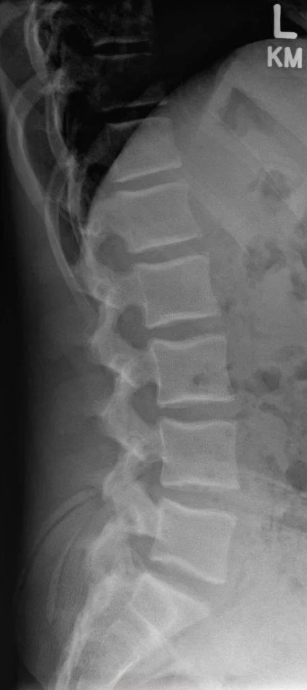
Lateral View
Taken from the side of the patient, with the shoulder positioned closest to the X-ray detector.
Helps visualize the disc spaces, vertebral body alignment, and the relationship between L5 and S1 from a sagittal (side) perspective.
Images need to be taken at neutral , flexion (bending forward) and extension (bending backward) positions.
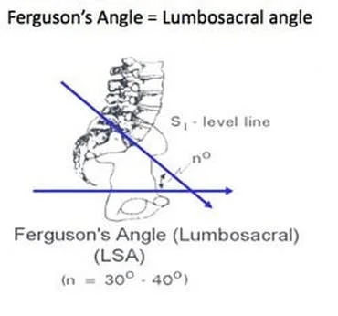
Ferguson View
Involves angling the X-ray beam cranially (upward) at 30–40 degrees while the patient remains in the AP position.
Enhances visualization of the lumbosacral junction, specifically the transverse processes of L5, and their articulation or fusion with the sacrum.
Image is taken while patient maintains a neutral posture.
References:
Alonzo, F., Cobar, A., Cahueque, M., & Prieto, J. A. (2018). Bertolotti’s syndrome: An underdiagnosed cause for lower back pain. Journal of Surgical Case Reports, 2018(10), rjy276. https://doi.org/10.1093/jscr/rjy276
Castellvi, A. E., Goldstein, L. A., & Chan, D. P. K. (1984). Lumbosacral Transitional Vertebrae and Their Relationship With Lumbar Extradural Defects. Spine, 9(5), 493.
Jenkins, A. L., O’Donnell, J., Chung, R. J., Jenkins, S., Hawks, C., Lazarus, D., McCaffrey, T., Terai, H., & Harvie, C. (2023). Redefining the Classification for Bertolotti Syndrome: Anatomical Findings in Lumbosacral Transitional Vertebrae Guide Treatment Selection. World Neurosurgery, 175, e303–e313. https://doi.org/10.1016/j.wneu.2023.03.077
Kapetanakis, S., Chaniotakis, C., Paraskevopoulos, C., & Pavlidis, P. (2017). An Unusual Case Report of Bertolotti’s Syndrome: Extraforaminal Stenosis and L5 Unilateral Root Compression (Castellvi Type III an LSTV). Journal of Orthopaedic Case Reports, 7(3), 9–12. https://doi.org/10.13107/jocr.2250-0685.782
McGrath, K., Schmidt, E., Rabah, N., Abubakr, M., & Steinmetz, M. (2021). Clinical assessment and management of Bertolotti Syndrome: A review of the literature. The Spine Journal, 21(8), 1286–1296. https://doi.org/10.1016/j.spinee.2021.02.023
Zhu, W., Ding, X., Zheng, J., Zeng, F., Zhang, F., Wu, X., Sun, Y., Ma, J., & Yin, M. (2023). A systematic review and bibliometric study of Bertolotti’s syndrome: Clinical characteristics and global trends.
X-Ray imaging sources:
AP view - Bickle I, Normal AP lumbar spine. Case study, Radiopaedia.org (Accessed on 30 Jun 2025) https://doi.org/10.53347/rID-46401
Lateral view - Bickle I, Normal lateral lumbar spine radiograph. Case study, Radiopaedia.org (Accessed on 30 Jun 2025) https://doi.org/10.53347/rID-46575
Ferguson view - https://chbradiography.weebly.com/ferguson-view.html