Identifying BSy anatomy using X-Ray
Proper identification and classification of lumbosacral transitional anatomy requires targeted X-ray imaging techniques. Physicians familiar with this condition typically use a combination of three imaging views and three positional postures to accurately visualize the anatomy and assess spinal mobility.
It is important to note that these views are not part of a typical X-Ray order requested by a doctor. To have these views be taken by the radiologist, your doctor must explicitly state in the X-Ray order to include all three of these imaging views and positional postures.
Recommended X-Ray imaging views
AP (Anteroposterior) View
Taken from the front of the patient, with the X-ray beam directed straight through the abdomen to the back.
Used to assess the alignment and width of the transverse processes and their relation to the sacrum.
Image is taken while patient maintains a neutral posture (standing upright with a natural spinal posture).

Lateral View
Taken from the side of the patient, with the shoulder positioned closest to the X-ray detector.
Helps visualize the disc spaces, vertebral body alignment, and the relationship between L5 and S1 from a sagittal (side) perspective.
Images need to be taken at neutral , flexion (bending forward) and extension (bending backward) positions.
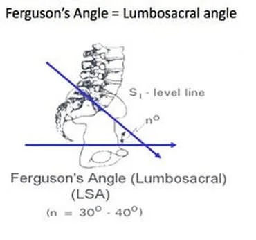
Ferguson View
Involves angling the X-ray beam cranially (upward) at 30–40 degrees while the patient remains in the AP position.
Enhances visualization of the lumbosacral junction, specifically the transverse processes of L5, and their articulation or fusion with the sacrum.
Image is taken while patient maintains a neutral posture.